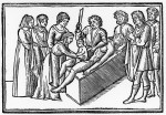
 Caesarean sections have the reputation of being named after Julius Caesar because he was delivered surgically from his mother Aurelia. That is a myth that grew from a misunderstanding of the ancient sources in the 10th century. Pliny states in Natural History (Book VII, Chapter 7) that the dictator’s branch of the Julius family acquired the cognomen of Caesar because the first to bear the name was excised from his mother’s womb (from the Latin “caedo,” meaning “to cut”). In other words, the Caesars were named after the section, not the other way around, and Julius just inherited the moniker. Anyway Aurelia lived for many years after her son’s birth, and c-sections were only performed on dead or dying women because an abdominal incision was considered impossible to survive at that time.
It was impossible for a very long time after Caesar, as a matter of fact. Without anesthesia, the pain from abdominal surgery would cause massive traumatic shock. If that didn’t kill you on the spot, the blood loss would. If by some miracle the mother managed to survive the immediate dangers, infection would surely finish the job. It wasn’t until the second half of the 19th century that the advances in surgical procedure — Listerism to combat infection, anesthetics like ether, nitrous oxide and chloroform to keep shock at bay — made caesarean sections a consistently survivable option for emergency delivery.
There are a few reports of women managing to survive c-sections from before modern surgery. The earliest dates to 1500 and was first included as a case history almost a century later in a 1581 obstetrics monograph, L’hysterotomotokie ou enfantement césarien, by Parisian physician Francois Rousset who named the procedure after Caesar. The mother was the wife of a swine-gelder named Jacob Nufer from Turgau, Switzerland. After days of labour and with both his wife and baby in mortal danger, Nufer appealed to the town council to let him try to operate on his wife since none of the local surgeons were willing to take the risk. Assisted by a midwife and a cutter of bladder stones, Nufer cut into his wife and delivered a living child abdominally. Mrs. Nufer not only survived, but went on to have four more children. There are some records backing up the story, but there is no specific reference to the uterus being cut, so some scholars believe it was a laparotomy for sudden, acute abdominal pain rather than a caesarean.
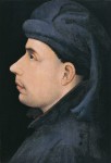
![]() Now obstetrician and medical historian Dr. Antonin Parizek of Charles University believes he may have found an earlier case: Beatrice of Bourbon, wife of the King of Bohemia John of Luxembourg who gave birth to their son in Prague in 1337. Beatrice was very young when she married the widowed king, just 14 years old, and it was not the happiest of matches. She had no interest in learning Czech of even German and was really only able to communicate with Blanche of Valois, the wife of her stepson (and future Holy Roman Emperor) Charles. King John spent all of two months at his court in Prague with his new bride in 1336, which was long enough to impregnate her. On February 25, 1337, Wenceslaus was born.
Now obstetrician and medical historian Dr. Antonin Parizek of Charles University believes he may have found an earlier case: Beatrice of Bourbon, wife of the King of Bohemia John of Luxembourg who gave birth to their son in Prague in 1337. Beatrice was very young when she married the widowed king, just 14 years old, and it was not the happiest of matches. She had no interest in learning Czech of even German and was really only able to communicate with Blanche of Valois, the wife of her stepson (and future Holy Roman Emperor) Charles. King John spent all of two months at his court in Prague with his new bride in 1336, which was long enough to impregnate her. On February 25, 1337, Wenceslaus was born.
The only contemporary records of his birth that have survived are two letters from Beatrice announcing the birth in Latin. They do not mention a c-section. In fact, she goes out of her way to say that her son was born “salva incolumitate nostri corporis,” an unusual phrasing which directly translates to “without breaching our body.” The health of the mother is almost never mentioned in comparable announcements from 14th century from members of the Luxembourg and Bourbon dynasties. The focus is on the child. It’s odd that she even went there. Parizek believes her insistence that her body was intact was a response to rumors of surgical intervention in a difficult birth. Beatrice had to yet to be crowned Queen of Bohemia, and there was a strong association of the bodies of rulers, chosen by God, as physical manifestations of their souls and as symbols of the church. A large slice across the gut through which the king’s son had had to be fished out did not fit the mold of sacred and inviolate royal bodies.
 It’s later sources that declare outright that Wenceslaus was born by c-section. The earliest is an early 15th century Flemish rhyming chronicle, Brabantsche Yeesten, by an anonymous author. There are verses in the chronicle that state that the boy was “taken from his mother’s body” and her wound healed. The author is amazed by this, because he had only ever heard of such an operation being performed on Julius Caesar. In 1549, Richard de Wassebourg, archdeacon of the Verdun Cathedral, reported the event in his book Antiquitez de la Gaule Belgique. At Wenceslaus birth, he said, “his mother Beatrice was opened up without dying.” Another reference is found in Mars Moravicus, written by Thomas Pesina of Čechorod, Vicar General and the Chapter Dean St. Vitus Cathedral in Prague, in 1677. Pesina states: “John had a son named Wenceslaus, taken from the queen Beatrice of Bourbon, or rather from the maternal womb, without endangering the mother, rarely such a lucky example of recovery [or healthy fertility].”
If Beatrice really did survive a caesarean in 1337, it was a perfect storm of very good luck for her and the future Duke of Luxembourg, her son.
Prague was a center of education, but also of the medical care of the royal family. In light of the health of John of Bohemia [– he suffered from ophthalmia and was basically blind by the time he married Beatrice –] a number of the most educated physicians of the time were present around the king. It can be presumed that they had the skills for the procedure, that is, cutting out a fetus from a dead or dying pregnant woman. Considering what we know, it can be rejected a priori that it was a case of deliberately saving the mother. In fact, the abdominal removal of the child from an apparently dead mother could partly explain the event described.
If that is indeed what happened, then with likelihood bordering on certainty Beatrice of Bourbon was considered to be dead when the procedure occurred. One explanation could be seizures as a complication of eclampsia. The cut would have had to be made immediately after the onset of such a state. The pain from the operation may have been the reason for a change in consciousness or awakening, and the stress reaction of the mother could also hypothetically explain why she did not bleed to death.
Suturing of wounds, especially of the abdominal wall, was a completely unknown procedure at that time. And that later complications did not arise from the non-sterile environment the operation was performed in push this hypothesis to the edge of reality. On the other hand, there is written evidence that using similar methods, with no anesthesia, surgical hemostasis or antiseptic conditions, the first experiments removing children through the abdomen after labor lasting several days were performed in the 17th to 19th centuries, and almost always in a home setting. It was very rare, but some women survived these operations.
Beatrice never had another child, which is another tick in the caesarean column, but she ended up outliving her first husband, who died in a famous charge against the English at the Battle of Crécy in 1346, by almost 40 years. Blind and 50 years old but still keen to fight, John tied his horse to his knights’ and rode fearlessly against the enemy. His was the only charge to break through the English lines, and the English killed them all. Beatrice also outlived all of her stepsons and even her son Wenceslaus, albeit only by 16 days.
You can read the whole fascinating paper published in the journal Ceska Gynekologie in English here. It touches on the history of c-sections, surgery, sutures, anesthetics and antiseptics, as well as on the specific case of Beatrice of Bourbon.
