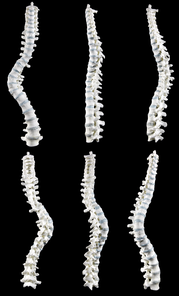
 For those of you who are over all this Richard III malarkey (hi anja!), I hope you understand why this post has to be. There’s a rotating spine gif here, people. How can I be expected to resist that? I’m only human. Besides, the question of Richard’s spinal deformity, its existence, nature and extent, has been the subject of many histories and even more theatrical performances for more than five centuries.
Now we have some real answers courtesy of the University of Leicester team which has published a brief paper on Richard’s spine in The Lancet. You can read it free of charge if you register on the site.
When a body decomposes, different parts break down at different rates. Ligaments that hold the spine together are some of the last ones to decompose, so usually the way the spine is found in the grave is how it was in life. The curvature in Richard’s spine could not have been a function of how he was placed. This was confirmed by examination of the bones, which found that the vertebrae of the curve are slightly different shapes and sizes. The only way those bones would fit together in life was in a spine with scoliosis.
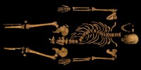
The skeleton laid out on a flat surface, however, only shows the sideways curvature of the spine. It takes a 3D model to see the full picture of the condition. The bones were scanned on a multi-detector CT scanner which takes high resolution images from every side, allowing them to be viewed as a whole 3D structure or in slices across any plane. The bones obviously were not joined, since the soft tissue is all gone and there is no software that will take the disconnected bones and put them back together the way they were in life. Usually that work is done by creating models.
 The team was able to use the imaging data to generate a model which was printed out in a polymer using the advanced 3D printing equipment of the Wolfson School of Mechanical & Manufacturing Engineering at Loughborough University in Leicestershire. This produces a near identical copy of the bones, only the model is durable, light weight and easily passed around, giving scientists the opportunity to study the skeletal structure without having to handle fragile human remains. Even after the king has been reburied, therefore, experts will still be able to examine his bones.
The bones of the spine join at three places: the gap between two vertebrae where there’s a disc and two facet joints at the back. With the plastic model, experts drilled a small hole in the center of each vertebra and ran a wire through them, separating each bone with a felt pad standing in for the disc. They then joined the facet joints using a similar technique. They saw that while the lumbar vertebrae in the lower spine appeared quite normal and fit together in a standard way, as they rose in the spine the osteoarthritic degeneration in the facet joints that was caused by the scoliosis increased markedly, deforming the joints. That deformity meant the bones fit together in a very specific way, an enforced thoracic curve that is the s-shaped bend in the spine we saw in the photographs of the skeleton in situ and in the lab. The measurement of the extent of the spinal curvature, called a Cobb angle, is 65-85 degrees. In today’s scoliosis patients that would be considered a large curvature to be corrected by the surgical implantation of metal rods. Once they reached the upper thoracic vertebrae, the facet joints returned to normal and the spine straightened out.
In addition to the sideways s-curve, the 3D model illuminates the spiral twist of the spine that you can only see when the spine is rotated. (You could see it even more clearly if the ribs were attached, but they haven’t 3D printed any ribs yet and probably won’t because many of them were broken when unearthed.) The model shows that the ribs on Richard’s back would have stuck out significantly on the right side, while they were sunken on the left. When he leaned forward, the prominent ribs on the right side of his back would have formed a hump. This would not have been visible, however, when he was clothed and in most any other position than leaning over, so all those pillows stuffed under costumes are way off.
The physical disfigurement from Richard’s scoliosis was probably slight since he had a well balanced curve. His trunk would have been short relative to the length of his limbs, and his right shoulder a little higher than the left. However, a good tailor and custom-made armour could have minimised the visual impact of this. A curve of 70—90° would not have caused impaired exercise tolerance from reduced lung capacity, and we identified no evidence that Richard would have walked with an overt limp, because the leg bones are symmetric and well formed.
He may or may not have had back pain. If his spinal curvature had been magically straightened, he’d have been 5'8" tall, about average for a man of the period. With the scoliosis he was two to three inches shorter.
The polymer model was photographed from 19 angles and the images used to create an interactive 3D model. You can click on it and drag it from side to side to examine the recreated spine from any perspective.